Penile Synovial Sarcoma: Clinical and Radiological findings
Sarcoma sinovial de pene: Hallazgos clínico radiológicos
DOI:
https://doi.org/10.25176/RFMH.v21i1.3090Abstract
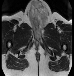
A 22-year-old man presented with a 6-month-old 8 cm hard tumor at the base of the penis, whose biopsy was consistent with synovial sarcoma (Figure 1). The magnetic resonance imaging (MRI) showed a tumor that compromises the base of the penis, the left pillar and partially the right pillar, extending to the distal 2/3, without ruling out urethral infiltration (Figure 1).
Downloads
Download data is not yet available.
Downloads
Published
2020-12-17
How to Cite
Espinoza-Figueroa, J., Uribe Rivera, A. K., & Luna-abanto, J. (2020). Penile Synovial Sarcoma: Clinical and Radiological findings: Sarcoma sinovial de pene: Hallazgos clínico radiológicos. Revista De La Facultad De Medicina Humana, 21(1). https://doi.org/10.25176/RFMH.v21i1.3090
Issue
Section
Photo Gallery
License
Copyright (c) 2020 Revista de la Facultad de Medicina Humana

This work is licensed under a Creative Commons Attribution 4.0 International License.